Electron microscopes
Throughout their development, the magnification of light microscopes has increased but very high magnifications are not possible. The maximum magnification with a light microscope is around √ó1500.
The limits of the light microscope
The magnification of a microscope is not the only factor that's important when viewing cells. The detail that can be seen is also important.
The ability to see greater detail in an image depends on the The fineness of detail that can be seen in an image - the higher the resolution of an image, the more detail it holds. In computing terms, resolution is measured in dots per inch (dpi).. This is the ability to see two points as two points, rather than merged into one.
Think about a digital photo. It can be enlarged, but over a certain size, you won't be able to see any more detail. It will just become blurry.
The resolution of a light microscope is around 0.2 μm which can also be written as 200 nm. This means that it cannot distinguish two points closer than 200 nm.
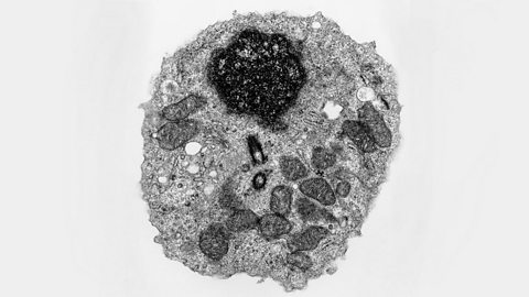
The electron microscope
Electron microscopes use a beam of electrons instead of light rays.
There are two types of electron microscope:
- The scanning electron microscope (SEM) has a large The distance between the nearest and farthest objects in focus. so can be used to examine the surface structure of specimens. SEMs are often used at lower magnifications.
- The transmission electron microscope (TEM) is used to examine thin slices or sections of cells or A group of similar cells that carry out the same function, eg muscle tissue..
TEMs have a maximum magnification of around x1,000,000, but images can be enlarged beyond that photographically. The limit of resolution of the transmission electron microscope is now less than 1 nm.
The TEM has revealed structures in cells that are not visible with the light microscope.