Ultrasound imaging
Ultrasound imaging creates a picture of something that cannot be seen directly, such as an unborn baby in the womb (a foetus), or faults and defects inside metals.
These uses rely on what happens when ultrasound waves meet the boundary between two different materials.
Medical uses of ultrasound
The best known example of the use of ultrasound is medical imaging, to 'see' inside a body.
An ultrasound scanner is simply run over the skin to obtain an image of what's inside.
The scanner probe has a built-in transducer that directs ultrasound pulses down into the body.
As the waves travel through the different bones and tissues, some are reflected off a boundary but some travel further to be reflected back up again off another boundary as some parts have different densities.
- some of the ultrasound waves are reflected at a boundary;
- the time taken for the waves to leave a source and return to the detector is measured;
- the depth of the boundary can be determined using distance = speed of sound in the material x the time taken.
The probe receives the reflected waves and a computer connected to the scanner uses them to draw an image on a screen.
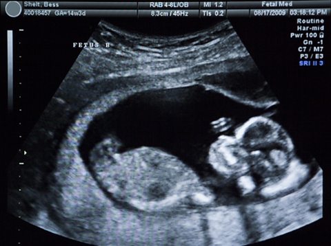
Scans of foetuses (unborn babies developing in the womb) are made this way and are used, for example, to measure the diameter of the head of a foetus so that growth can be monitored.
Ultrasound imaging also helps to diagnose problems with the:
- heart;
- kidneys;
- blood vessels;
- bladder.
The best known non-imaging medical use of ultrasound is the breaking of kidney stones.
The vibrations caused by the ultrasound shake apart the kidney stones, breaking them up.
Advantages of using ultrasound in medicine
- Ultrasound waves pass through tissue without causing harm, unlike x-rays which can damage DNA inside cells.
- Ultrasound equipment is relatively cheap, portable and easy to use.
- Images of internal organs can be seen without having to operate on patients.